Sunday, September 16, 2012

PET/CT

http://www.asiancancer.com/technology-equipment/111.html   PET/CT: A technology in anatomical form to capture the images of function, metabolism and receptor
  Scouts of tumor with more than 90% accurate diagnostic rate
  What is PET/CT?
  As a most high-end medical imaging devise in the world, PET/CT is currently the only technology in anatomic form to obtain images of function, metabolism and acceptor.
  Featuring no wounds, it detects the minimum cancer focus and discovers potential cancerous focus early of high resolution and sensitivity. With accurate diagnostic rate of more than 90%, PET/CT plays an important role in planning treatments.
  Principle
  Tumor cell’s uncontrolled proliferation, which including increased DNA synthesis and raised consumption of metabolic substrates like amino acids and glucose, is a remarkable difference from normal tissue cells metabolism. Via tracing and labeling metabolic substrates or analogs by radionuclide like 18F-FDG(18F-2-fluro-D-deoxy-glucose), tumor metabolism can be depicted in images directly. Those images can precisely and actively reflect the difference of cell metabolism between tumor tissue and body normal tissue, and then early diagnosis, planning treatments and monitoring curative effect could be done accordingly.
  Procedures for PET-CT
  Before the exam, the patient fasts and forbids glucose transfusion for greater than 6 hours; meanwhile avoids doing strenuous or long-time exercise.
  Nurse introduces exam procedures, measures patient’s height and weight, and then finishes the exam result.
  Doctor talks to patient about his/her medical history, checks patent’s blood glucose, explains to patient about informed consent before patient or his/her family sign it.
  Patient takes a rest for 45 minutes after PDG injection.
  Patient keeps quiet, walks less and avoids talking to others as possible; he / she also have to urine completely before CET/PT.
  PET-CT takes 30 minutes.
  In order to finish the PET-CT for whole-body, 2.5-3 hours should be allowed in advance.
  Advantages
  Safe and no wounds: Radionuclide for PET lingers in candidate’s body so short that no harm would do to the human body.
  High accuracy on diagnosing tumor benignancy, malignancy and staging: PET/CT can indicate tumor malignant degree while accurately detect the distant metastasis to lymph nodes.
  PET/CT can cut done the treatment costs: general treatment costs could be cut down by inchoate discovery and inchoate cure of tumor or via PET/CT to obtain more precisely diagnosis to perform effective therapy

Saturday, September 15, 2012

Interventional Therapy---Starve Tumor to Death


 Interventional therapy is a mini-invasive therapy that under the guidance of medical image equipment. It mainly has two categories: vascular interventional therapy and non-vascular interventional therapy or diagnosis. With a 1-2 millimeter cut, paracentesis could be performed with the lead of medical image equipment like CT. Then the special tube, transend or other sophisticated apparatus are introduced into human body for diagnosing the conditions or draw some tissues for topical therapy at the same time or any other time. Interventional therapy is applied vastly in tumor diagnosis and treatments because of its advantages like no operation, small wounds, fast recovery and good effect.

PDT (Photodynamic Therapy) -- the "Photon Bullets" of Anti-cancer


Photodynamic therapy is a non-invasive therapy using photosensitizer and laser to effectively destroy cancer cells. PDT was approved by the U.S. FDA for clinical application in 1996 and approved for clinical application by Chinese SFDA in 2003.

  Cancer tissue absorbs a great deal of photosensitizer injected intravenously whereas normal tissue absorbs little. Then special laser irradiation is employed to kill tumor. During the process, photosensitizer interacts with cancer cell and leads to intoxication through necrosis of cancer cells on photodynamic effects, thus the tumor gradually disappears.

ture=plcp

What Tests Can Be Done to Detect Colon Cancer


“What tests can be done to detect colon cancer?’’ people would ask such question when they are involved in colon cancer or suspected symptoms. When colon cancer is suspected, either a lower GI series (barium enema x-ray) or colonoscopy is performed to confirm the diagnosis and to localize the tumor.
Tests that can be done to detect colon cancer:
1. Barium enema involves taking x-rays: According to experts in Modern Cancer Hospital Guangzhou, a barium enema involves taking x-rays of the colon and the rectum after the patient is given an enema with white and chalky liquid containing barium. The barium outlines the large intestines to the x-rays. Tumors and other abnormalities appear as dark shadows on the x-rays.
 2. Colonoscopy: “Colonoscopy is a procedure whereby a doctor inserts a long, flexible viewing tube into the rectum for the purpose of inspecting the inside of the entire colon,” said Prof Peng Xiaochi, an oncology expert from Modern Cancer Hospital. Colonoscopy is generally considered more accurate than barium enema x-rays, especially in detecting small polyps. If colon polyps are found, they are usually removed through the colonoscopy and sent to the pathologist. The pathologist examines the polyps under the microscope to check for cancer. While the majority of the polyps removed through the colonoscopies are benign, many are precancerous. Removal of precancerous polyps prevents the future development of colon cancer from these polyps.
3. Biopsy: If cancerous growths are found during colonoscopy, small tissue samples (biopsies) can be obtained and examined under the microscope to confirm the diagnosis. If colon cancer is confirmed by a biopsy, staging examinations are performed to determine whether the cancer has already spread to other organs. Since colorectal cancer tends to spread to the lungs and the liver, staging tests usually include chest x-rays, ultrasonography, or a CAT scan of the lungs, liver, and abdomen.
4. Blood test: Sometimes, doctors in Modern Cancer Hospital Guangzhou may obtain a blood test for CEA (carcinoembyonic antigen). CEA is a substance produced by some cancer cells. It is sometimes found in high levels in patients with colorectal cancer, especially when the disease has spread.


Thursday, September 13, 2012

Colon Cancer


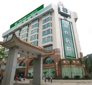
Colon cancer: A malignancy that arises from the inner lining of the colon. Most, if not all, of these cancers develop from colonic polyps. Removal of these precancerous polyps can prevent colon cancer, according to experts from Modern Cancer Hospital Guangzhou.

The colon is the part of the digestive system where the waste material is stored. The rectum is the end of the colon adjacent to the anus. Together, they form a long, muscular tube called the large intestine (also known as the large bowel). Tumors of the colon and rectum are growths arising from the inner wall of the large intestine. Benign tumors of the large intestine are called polyps. Malignant tumors of the large intestine are called cancers.

Oncology experts from Modern Cancer Hospital Guangzhou stated that colon polyps and early colon cancers cause no signs or symptoms. Full-blown colon cancer can cause occult (a microscopic amount of) blood in the stool, overt rectal bleeding, bowel obstruction, and weight loss.

Risk factors for colon cancer include a family history of it or of colonic polyps, and long standing ulcerative colitis. The overall risk can be reduced by following a diet low in fat and high in fiber.
Colon cancer is preventable and curable. It is preventable by removing the precancerous colon polyps. It is curable if detected early when it can be surgically removed before it has spread to other parts of the body. If screening and surveillance programs were practiced universally, there would be a tremendous reduction in the incidence and mortality of colon cancer.

Globally, cancer of the colon and rectum is the third leading cause of cancer in males and the fourth leading cause of cancer in females. The frequency of colorectal cancer varies around the world. It is common in the Western world and is rare in Asia and Africa. In countries where the people have adopted western diets, the incidence of colorectal cancer is increasing.